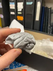
Chinese hamster ovary cells
Description
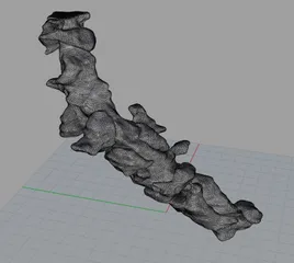
PDF2 dividing wild-type Chinese hamster ovary (CHO) cells reconstructed from a scanning electron microscope data, 4000x magnification. The cell membrane on these is bubbly- packed with 'blebs'. Scanning electron microscope images by Jess Holz, 3D reconstruction by Ahmadreza Baghaie in the laboratory of Dr. Zeyun Yu, UW-Wisconsin Milwaukee Computer Science Department: https://pantherfile.uwm.edu/yuz/www/bmv/index.html
How I Designed This
3D reconstruction from scanning electron microscope data
Created by 3d reconstruction from a stereo-pair of scanning electron microscope images, taken at about 8 degrees tilt relative to each other. The algorithm creates a dense reconstruction in a manner not entirely unlike 123d catch. The algorithm was developed by the laboratory of Dr. Zeyun Yu, University of Wisconsin-Milwaukee Computer Science Department. See our latest paper by Tafti et al: 3DSEM++: Adaptive and intelligent 3D SEM surface reconstruction, available here: http://www.sciencedirect.com/science/article/pii/S0968432816300750

Original scanning electron microscopy data by Jess Holz

3d reconstruction.
Category: BiologyTags
Model origin
The author marked this model as their own original creation. Imported from Thingiverse.


